Adele Diamond: A Pioneer in Developmental Cognitive Neuroscience

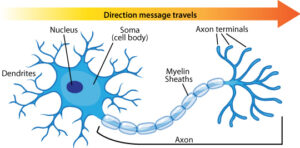
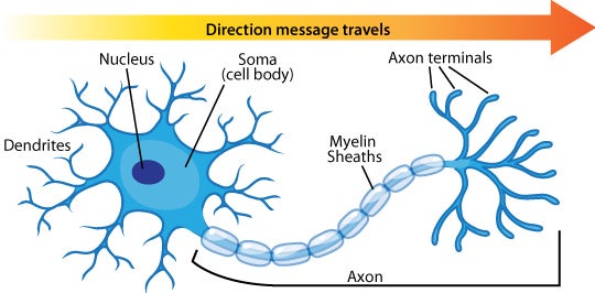
Adele Diamond is a developmental cognitive neuroscientist. She is currently a professor of Developmental Cognitive Neuroscience at the University of British Columbia. She is also a Fellow of the Royal Society of Canada. Diamond has recently been named one of the 15 most influential neuroscientists. Diamond’s main area of research is executive functions. Executive functions include many different aspects; cognitive flexibility, working memory, inhibitory control, and attentional shifting. Executive functions are important for creative and flexible problem-solving, meeting unanticipated challenges, self-control, reasoning, and the discipline to persevere. Diamond specifically looks at how executive functions are affected by biological and environmental factors in children. Her research has improved treatment for medical disorders and ADHD, as well as impacted early education. Diamond is now looking into the possible roles of traditional activities like music and dance in the improvement of executive functions, academic outcomes, and mental health.

When Diamond began her research in the 1980s on developmental cognitive neuroscience, it was a major landmark in the combination of developmental psychology, cognitive science, and neuroscience. In the 1990s, Diamond and colleagues made discoveries that led to the improvements of the treatment for phenylketonuria (PKU). They determined the biological mechanism that causes executive function deficits in children who are being treated for PKU. In the 2000s, Diamond conducted research on the clinical differences between ADHD with hyperactivity and inattentive-type ADHD. Her research was greatly appreciated by ADHD patients. She also conducted research on the effects of education on executive functions. She discovered that the better the child’s executive functions, the better their performance on standardized measures of academic performance. This research found that executive functions could be improved in children by teachers in classrooms, without computerized training. Diamond’s research has led to an increase in interest in the capability of early interventions to improve executive functions to combat mental health issues and school issues.

Vilayanur Ramachandran: Phantom Limbs and Neural Plasticity

Vilayanur S. Ramachandran (1951-) is a neuroscientist from Tamil Nadu, India. He is best known for his work in behavioral neurology and visual psychophysics. He is currently a professor in the Department of Psychology and the Graduate Program in Neurosciences at the University of California, San Diego, and he is the director of the Center for Brain and Cognition. Ramachandran studied at the University of Madras in Chennai, India, as well as Trinity College at the University of Cambridge. Ramachandran has conducted research on a variety of topics. He began by doing research on human visual perception. He then moved on researching neurological syndromes like phantom limbs, body integrity identity disorder, and the Capgras delusion. Ramachandran also worked with the understanding of synesthesia and invented the mirror box. Ramachandran is known for using simpler technology in his experiments.
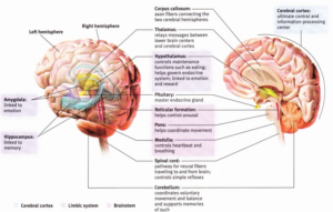
Ramachandran’s Theories:

Phantom Limbs: Ramachandran theorized that there was a connection between phantom limbs and neural plasticity in the adult human brain.

Mirror Visual Feedback/Mirror Therapy: Ramachandran invented the mirror box and presented mirror visual feedback as a treatment for phantom limb paralysis.

Neural Cross-Wiring/Synesthesia: Ramachandran was one of the first scientists to theorize that grapheme-color synesthesia comes from a cross-activation between brain regions.

Mirror Neurons: Ramachandran advocates for the importance of mirror neurons. Ramachandran went so far as to say, “Mirror neurons will do for psychology what DNA did for biology…” He believes that mirror neurons play a role in empathy, imitation learning, language development, and self-awareness.

“Broken Mirrors: Theory of Autism: Ramachandran hypothesized that a loss of mirror neurons might be the main deficit that explains many of the symptoms and signs of autism spectrum disorders. This hypothesis is controversial because the research did involve measuring mirror neuron activity directly, and instead showed that children with ASD displayed abnormal EEG responses when they observed the activities of other people.

Xenomelia (Apotemnophilia): Ramachandran theorized that apotemnophilia is a neurological disorder caused by damage to the right parietal lobe of the brain.

https://www.theguardian.com/theobserver/2011/jan/30/observer-profile-vs-ramachandran

Brain Structures and their Function

The human brain is made up of many different parts that all have their own unique function. They work together to accomplish all functions of our lives, from memories to actions. When a part of the brain does not work properly, the consequences can be severe. With an understanding of what each part of the brain does, one can start to understand how humans function.

One of the most important parts of the brain is the cerebral cortex because it is what makes us human. It is the ultimate control and information-processing center of the brain. The cerebral cortex is the outer layer of neural tissues. This tissue plays a key role in memory, attention, perception, awareness, thought, language, and consciousness.

The Brain Structure and their Functions Credit: http://owlriginal.blogspot.com/2014/01/brain-regions-part1.html#.WCVE8vkrLIU

A key component of the brain is to communicate messages between neurons. To accomplish this the two hemispheres of the brain must pass messages between each other. The axon fibers that are connecting the two cerebral hemispheres together is known as the Corpus Callosum. This neural tissue facilitates communication between the two sides of the brain.

The cerebellum enabled nonverbal learning and skill memory. It is also helps us judge time module our emotions and discriminate sounds and textures. Whereas the hippocampus helps process explicit memories and helps in the storage of memories.

A very important part of the limbic system is the amygdala. It is linked to emotions, especially aggression and fear. Along with the amygdala, the hypothalamus is also linked to emotion and reward. It also controls functions, such as eating.

Additional Resources

The Functions of the Four Lobes of the Brain


Human brain in x-ray view
Click to see the interactive map of the brain Credit:http://www.cbc.ca/news2/interactives/brain/

Our brains are made up of two hemispheres, the right and the left. Covering these hemispheres, is a thin surface layer of interconnected neural cells called the cerebral cortex. It is your brains thinking crown, your body’s ultimate control and information-processing center. The cerebral cortex is divided into four sections, or lobes. They include the frontal lobe, parietal lobe, occipital lobe, and temporal lobe.

The Four Lobes Credit: http://www.internetdict.com/wp-content/uploads/related_images/2016/01/14/what-are-the-four-lobes-of-the-brain_1.jpg

The frontal lobe is the portion of the cerebral cortex lying just behind the forehead. It is involved in speaking and muscle movements, as well as making plans and judgments. This area is best described as the control center of the brain.

Next, the parietal lobe is the portion of the cerebral cortex lying at the top of the head and toward the rear. This receives sensory input for touch and body position.

The Occipital lobe is the portion of the cerebral cortex lying at the back of the head. This area receives information from the visual fields and is associated with vision processing.

The forth lobe, the temporal lobe, is the portion of the cerebral cortex laying roughly above the ears. This includes the auditory areas, each receiving information primarily from the opposite ear.

Additional Information

Neurons: How do they work?

Neurons transmit messages

Our brain’s are made up of billions of nerve cells called neurons. These are the basic building blocks of the nervous system. To understand how our thoughts and actions, our memories and moods, we need to know how neurons work and communicate.

In 1873, Italian physician and scientist Camillo Golgi discovered a silver staining technique that is used to visualize nervous tissues under a light microscope. The method was named Golgi’s method. This was the start of studying neurons. Today, we now known more about the complexity of a neuron.

Although neurons can differ, they all consists of a cell body, dendrites, axon, and myelin sheaths.  The cell body directs all activities of the neuron.  This is connected to the bushy dendrite fibers that receive information, like messages, from other neurons and relay those messages toward the cell body. From there, the cell’s single axon fiber passes the message through its terminal branches from the cell body to the dendrites of other neurons or to muscles. In short, dendrites listen where as axons speak.

Neuron Diagram

Dendrites are shorter in length where as the axons can be very long, even several feet through the body. The axons are encased in a myelin sheath, a layer of fatty tissue that insulates them and speeds up their impulses. Every moment, messages are being sent back and forth at amazing speeds from neuron to neuron.

How synapse work

A neuron communicates with other neurons at a synapse. To send messages, a neuron releases chemical messengers, called neurotransmitters. Within 1/10,000 of a second, the neurotransmitter molecules crosses the synaptic gap and bind to a receptor sites on the receiving neuron.  When neurotransmitters attach to these receptors, they cause changes inside the receiving neuron and the message is delivered.This is best visualized as a key fitting into a lock. The excess neurotransmitters then drift away, are broken down by enzymes or are reabsorbed by the sending neurons, a process called re-uptake.

Understanding neurons helps us understand how thought, memories, actions, and moods are created. They even help explain how drugs effect our brains and create addictions. The more you know about the neuron, the easier it is to explain how our brain works.

Videos

Sites


Phineas Gage


In September 1848, Phineas Gage was working as a foreman of a crew cutting a railroad bed in Cavendish, Vermont. He was 25 years old.  It was on September 13, when he was using a tamping iron to pack explosive power into a hole when the powder detonated. The tempting iron, that was 43 inches (3 feet 7 inches) long, 1.25 inches in diameter and weighing 13.50 pounds, shot upwards and penetrated point first through Gage’s left cheek.  It ripped into his brain and exited through his skull. The tamping iron landed about 25 yards away from him.  Although this may have taken Gage’s sight from his left eye, he might not have lost consciousness. He was taken to be treated by Doctor John Martyn Harlow.

Phineas Gage’s Skull

John Harlow treated Gage for months after the accident and made observations about Gage’s health and behaviors. He state,d that the balance between his “intellectual faculties and animal propensities” seemed gone. He could no longer stick to plans, uttered profanity and showed little deference for his fellows.  Gage’s friends found him to be “no longer Gage,” after the accident.

Phineas Gage with the tamping iron in hand

After being refused his former job as a foreman, he went to work in a stable in New Hampshire, drove coaches in Chile, until he eventually joined relatives in San Francisco. This is where he died 12 years after the accident, in May 1860, at the age of 36, after a series of epileptic seizures.

Phineas Gage became one of the most famous patients in the area of neuroscience because his case was the first to suggest a link between brain trauma and personality changes.

Videos:

Sites: